The Human Heart: The Engine of Life
Introduction
 The heart stands as one of the most vital organs in the human body, serving as the tireless engine of our circulatory system. This extraordinary muscular pump beats approximately 100,000 times each day, propelling roughly 2,000 gallons of blood through an intricate network of vessels spanning 60,000 miles. From the first flutter in the womb until our final breath, the heart works ceaselessly, delivering life-sustaining oxygen and nutrients to every cell while efficiently removing metabolic waste. Understanding this remarkable organ—its complex anatomy, sophisticated function, and the diseases that threaten it—proves essential for maintaining cardiovascular health throughout our lives.
Main Article
The human heart represents an extraordinary achievement of biological engineering, functioning as a sophisticated dual-pump system that sustains life through continuous, precisely coordinated contractions. This remarkable organ has captivated scientists, physicians, and philosophers throughout history, serving not merely as a biological necessity but as a profound symbol of life, emotion, and human vitality across virtually every culture worldwide.
The heart's fundamental responsibility centers on circulating blood through two interconnected yet distinct pathways. The pulmonary circulation sends oxygen-depleted blood to the lungs for gas exchange, where carbon dioxide is released and fresh oxygen absorbed. The systemic circulation then distributes this newly oxygenated blood throughout the entire body, reaching every organ, tissue, and individual cell. This elegant dual-circuit design ensures optimal oxygen delivery while efficiently managing waste removal, creating a self-sustaining system of remarkable efficiency and endurance.
With the heart serving as its central command, the cardiovascular system comprises an astonishingly complex network of blood vessels that, if laid end to end, would stretch approximately 60,000 miles—enough to circle the Earth more than twice. The heart pumps between 1,500 and 2,000 gallons of blood daily, yet demonstrates extraordinary adaptability, adjusting its output moment by moment based on the body's ever-changing demands. During intense physical activity, cardiac output can increase fivefold or more, showcasing remarkable responsiveness and reserve capacity.
 The heart's electrical system orchestrates each heartbeat with split-second precision, ensuring perfect synchronization between chambers. The sinoatrial node, a specialized cluster of cells in the right atrium's upper region, serves as the heart's natural pacemaker, spontaneously generating electrical impulses 60 to 100 times per minute under resting conditions. These electrical signals propagate through specialized conduction pathways, coordinating atrial contraction first to fill the ventricles, followed milliseconds later by powerful ventricular contraction that propels blood into the great arteries. This intricate electrical system functions autonomously, requiring no conscious thought or control.
Blood flows through the heart in an invariable, one-way pathway maintained by four precisely engineered valves. Deoxygenated blood returning from bodily tissues enters the right atrium through the superior vena cava and inferior vena cava. From the right atrium, blood passes through the tricuspid valve into the right ventricle, which contracts to pump blood through the pulmonary valve into the pulmonary arteries. These arteries carry blood to the lungs, where it releases carbon dioxide and absorbs oxygen through the thin walls of millions of tiny air sacs called alveoli.
Freshly oxygenated blood returns from the lungs through four pulmonary veins, entering the left atrium. It then flows through the mitral valve into the left ventricle, the heart's most powerful chamber, which features walls three times thicker than the right ventricle. When the left ventricle contracts, it generates tremendous pressure, forcing blood through the aortic valve into the aorta, the body's largest and most important artery. From this central vessel, blood branches into progressively smaller arteries, arterioles, and eventually microscopic capillaries where nutrient and gas exchange occurs at the cellular level throughout every tissue in the body.
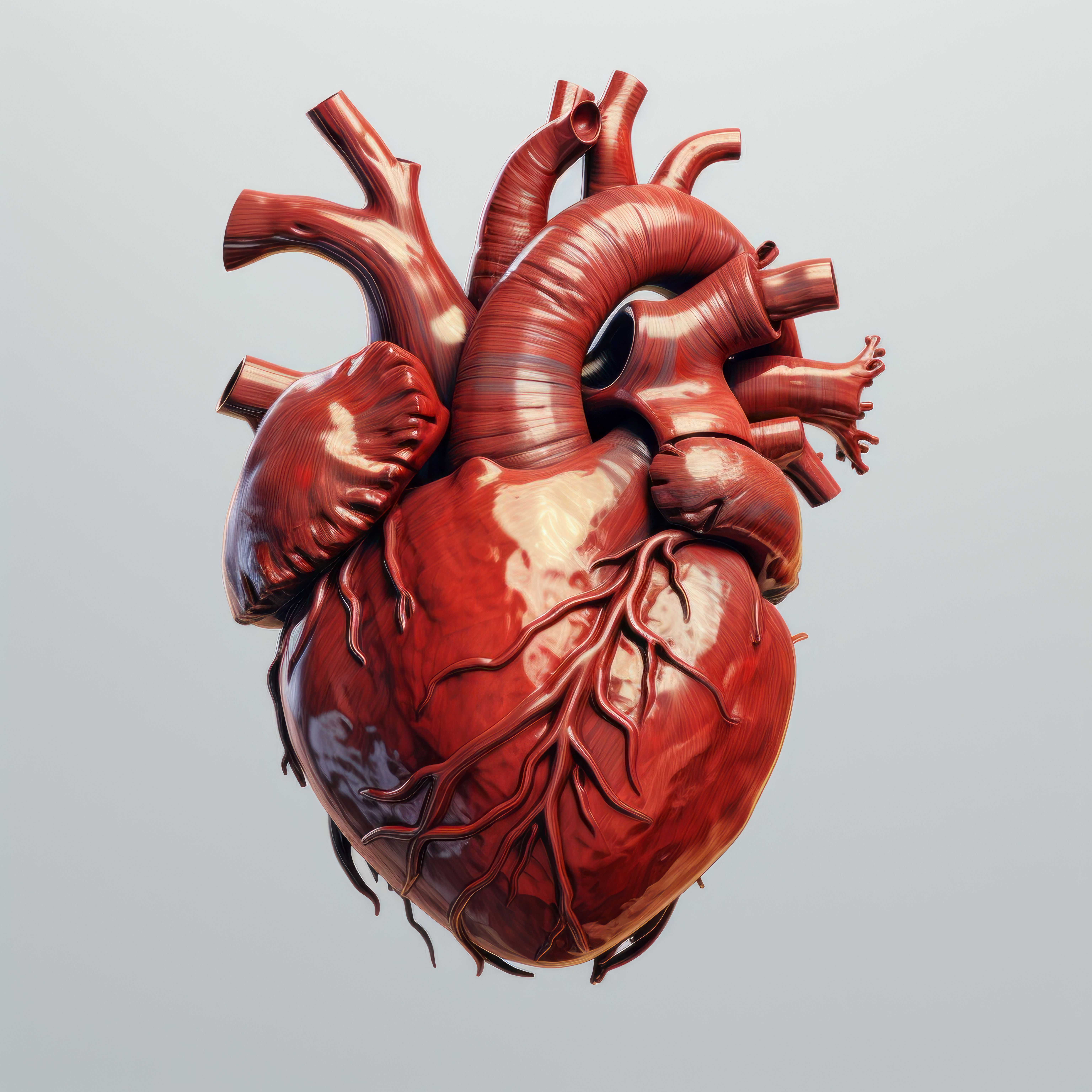
Remarkably, despite being filled with blood, the heart muscle itself requires its own dedicated blood supply, provided by the coronary arteries. These vital vessels branch directly from the aorta's base and course across the heart's surface like a crown. The left main coronary artery divides into the left anterior descending artery, which supplies the front and bottom of the left ventricle, and the circumflex artery, which wraps around to supply the left atrium and the left ventricle's side and back. The right coronary artery supplies the right ventricle, right atrium, and portions of the left ventricle's inferior wall. Any obstruction in these critical vessels can precipitate devastating consequences, including heart attack and potentially fatal cardiac events.
The heart plays a crucial role in blood pressure regulation through sophisticated interactions with nervous and endocrine systems. The autonomic nervous system exerts continuous influence over cardiac function: the sympathetic division accelerates heart rate and strengthens contractions during stress, physical exertion, or exercise, while the parasympathetic division slows the heart and promotes rest, recovery, and relaxation. Beyond its mechanical function, the heart produces natriuretic peptides, hormones that help regulate blood volume, sodium balance, and vascular tone, demonstrating that this organ functions not merely as a pump but as an integrated endocrine organ essential for overall cardiovascular health.
 Heart sounds—the familiar "lub-dub" audible through a stethoscope—provide valuable diagnostic information about heart function and health. The first heart sound results from closure of the atrioventricular valves (tricuspid and mitral) as ventricular contraction begins. The second sound occurs when the semilunar valves (pulmonary and aortic) snap shut as the ventricles relax and prepare for the next filling phase. Abnormal sounds, called murmurs, can indicate valve dysfunction, structural abnormalities, or turbulent blood flow patterns requiring careful medical attention and evaluation.
The heart demonstrates extraordinary adaptability throughout the human lifespan. During embryonic development, the heart emerges as one of the first functional organs, beginning to beat just 22 days after conception, well before most other organs have formed. As we age, the heart undergoes various structural and functional changes, including slight wall thickening, modest valve calcification, and decreased maximum achievable heart rate during intense exercise. However, these age-related changes need not significantly impair function, especially when supported by healthy lifestyle practices consistently maintained throughout life.
Function
The heart's primary function centers on pumping blood continuously throughout the body, maintaining vital circulation that delivers oxygen and nutrients to every cell while simultaneously removing carbon dioxide and metabolic waste products. This muscular organ contracts rhythmically and automatically, creating the precise pressure gradients necessary to propel blood through an extensive network of arteries, capillaries, and veins.
The cardiac cycle encompasses two fundamental phases that alternate throughout life. Systole represents the active contraction phase, during which the ventricles forcefully eject blood—the right ventricle sending deoxygenated blood to the lungs while the left ventricle propels oxygen-rich blood throughout the body via the aorta. Diastole constitutes the relaxation and filling phase, when the heart chambers expand passively to receive incoming blood from the veins. This alternating pattern typically occurs 60 to 100 times per minute in healthy resting adults, though this rate varies based on age, fitness level, and immediate physiological demands.
The heart possesses an intrinsic ability to regulate its own rhythm through a sophisticated electrical conduction system independent of conscious control. The sinoatrial node spontaneously generates electrical impulses that propagate across both atria, triggering their synchronized contraction. These signals then converge at the atrioventricular node, which introduces a brief delay allowing complete ventricular filling before transmitting the impulse through the bundle of His and Purkinje fiber network, ultimately triggering powerful ventricular contraction. This electrical choreography occurs with split-second precision, coordinating the mechanical events of each heartbeat.
Structural Organization
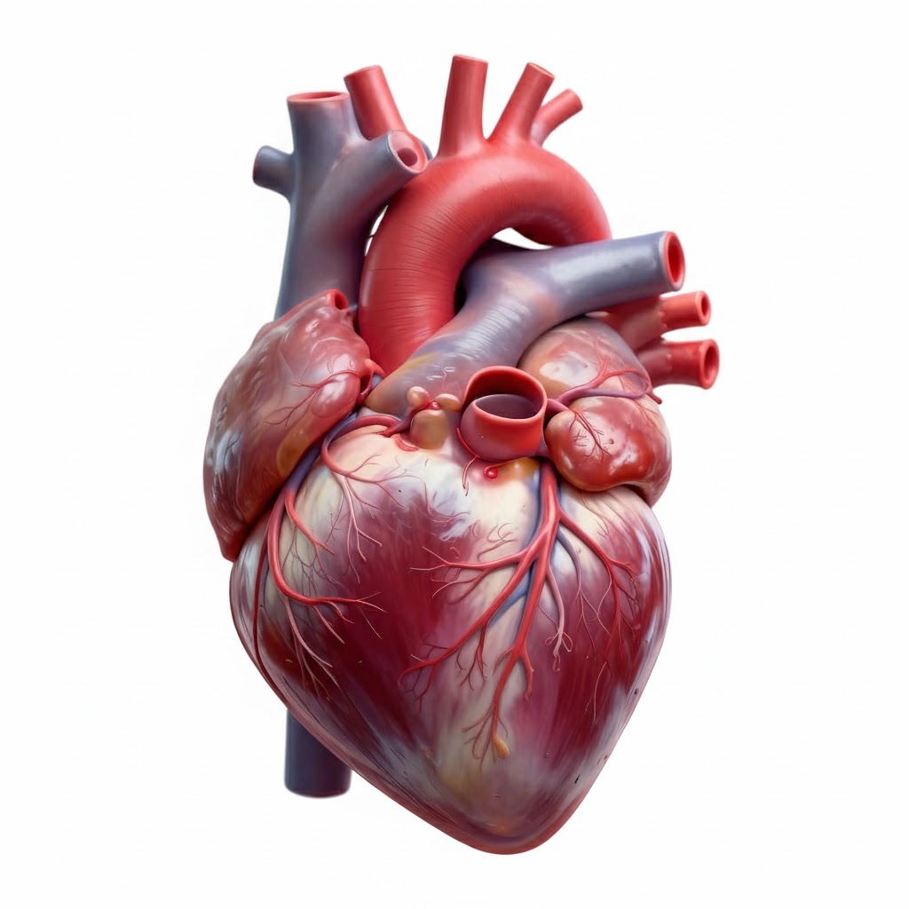
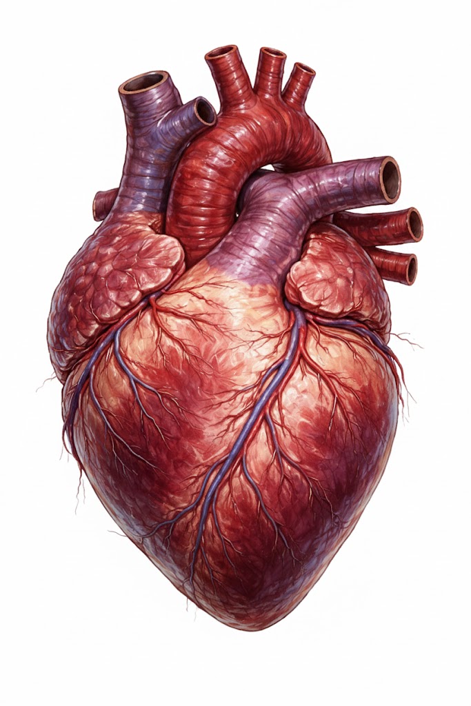
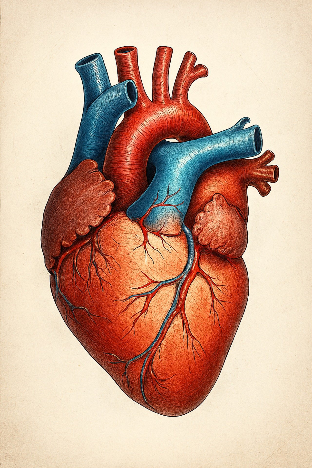
The heart's sophisticated structure directly reflects and enables its function as an efficient, powerful dual-pump system. Anatomically shaped like an inverted, slightly flattened cone, the heart consists primarily of specialized cardiac muscle tissue called myocardium. This unique muscle type possesses extraordinary properties enabling continuous rhythmic contractions without fatigue. The heart wall comprises three distinct layers: the endocardium, a smooth inner lining that prevents blood clotting; the myocardium, the thick muscular middle layer generating contractile force; and the epicardium, a protective outer layer containing coronary vessels.
Surrounding the entire organ, the pericardium forms a tough, double-layered protective sac that anchors the heart within the mediastinum while preventing excessive movement during vigorous activity. The fibrous pericardium forms the outer layer, while the serous pericardium consists of parietal and visceral layers. The narrow pericardial cavity between these layers contains lubricating serous fluid that virtually eliminates friction during the heart's ceaseless beating, allowing smooth cardiac motion throughout billions of contractions over a lifetime.
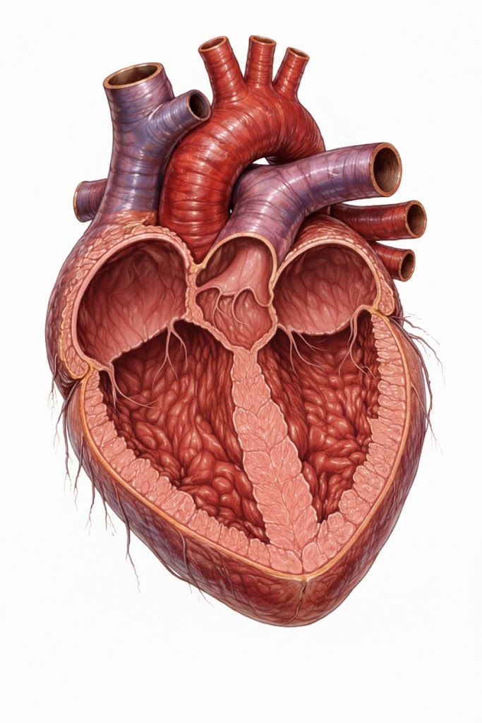
Four distinct chambers organize the heart's internal architecture into a synchronized dual-pump system. The right atrium receives deoxygenated blood returning from systemic circulation, while the right ventricle pumps this blood to the lungs for oxygenation. The left atrium receives freshly oxygenated blood from the pulmonary veins, and the powerful left ventricle propels this oxygen-rich blood throughout the entire systemic circulation.
Thick muscular partitions maintain complete separation between the heart's sides. The interventricular septum divides the left and right ventricles, while the interatrial septum separates the atria, preventing any mixing of oxygenated and deoxygenated blood and preserving system efficiency.
Four precisely engineered valves ensure strictly unidirectional blood flow through the heart. The tricuspid and mitral valves control flow between atria and ventricles, while the pulmonary and aortic valves regulate outflow from ventricles to great arteries. These valves open and close passively in response to pressure changes during the cardiac cycle, preventing backflow and maintaining forward blood progression.
Location and Shape
The heart occupies a protected position within the thoracic cavity, specifically residing in the mediastinum—the central compartment situated between the two lungs. Positioned slightly left of the body's vertical midline, approximately two-thirds of the heart's mass lies left of center. The heart rests behind the protective sternum (breastbone) and sits atop the diaphragm, the muscular sheet that separates the thoracic and abdominal cavities. This strategic placement shields the heart from external trauma while positioning it centrally for optimal blood distribution to all regions of the body.
The heart's orientation within the chest resembles a tilted cone that has toppled onto its side. The base, representing the broader posterior surface, faces toward the spine and slightly upward toward the right shoulder. The apex—the heart's pointed inferior tip—angles downward, forward, and distinctly leftward, typically positioned at the fifth intercostal space near the midclavicular line. This specific positioning explains why heartbeats are most prominently felt on the chest's left side.
Regarding dimensions, the average adult heart approximates the size of its owner's closed fist, measuring roughly 12 centimeters long, 8 centimeters wide, and 6 centimeters thick. A typical adult heart weighs between 250 and 350 grams, with male hearts generally larger than female hearts due to body size differences and hormonal influences. Despite its relatively modest size, this compact organ possesses the strength and endurance to sustain circulation throughout an entire lifetime.
Diseases
Cardiovascular diseases constitute the leading cause of mortality worldwide, claiming millions of lives annually and affecting countless others with chronic debilitating conditions. Coronary artery disease represents the most prevalent form, developing when atherosclerotic plaque accumulates within coronary arteries, progressively narrowing these critical vessels and restricting blood flow to the heart muscle. This insidious process often advances silently over decades before producing symptoms.
 Myocardial infarction, commonly termed a heart attack, occurs when coronary artery blockage becomes complete, depriving heart muscle tissue of oxygen and causing irreversible cell death. Symptoms typically include crushing chest pain or pressure, shortness of breath, arm or jaw pain, nausea, and profuse sweating, though presentations vary considerably, particularly in women and diabetics who may experience more subtle warning signs.
Heart failure describes a chronic condition wherein the heart cannot pump blood efficiently enough to meet the body's metabolic demands, resulting in fluid accumulation, profound fatigue, and exercise intolerance. This syndrome can result from various causes including prior heart attack, chronic hypertension, or valvular disease, and represents a growing public health challenge.
Cardiac arrhythmias encompass abnormal heart rhythms ranging from benign premature beats to life-threatening ventricular fibrillation. Valvular heart diseases occur when valves fail to open fully, called stenosis, or close completely, known as regurgitation, compromising circulation efficiency. Congenital heart defects represent structural abnormalities present from birth and vary widely in severity. Major risk factors for cardiovascular disease include hypertension, elevated cholesterol, diabetes, obesity, tobacco use, physical inactivity, poor dietary habits, chronic stress, and family history—many of which can be modified through lifestyle changes and medical intervention.
How to Protect Your Heart
Protecting cardiovascular health demands a comprehensive approach combining lifestyle modifications with proactive medical care. Regular physical activity represents perhaps the single most powerful protective strategy available. Adults should accumulate at least 150 minutes of moderate-intensity aerobic exercise weekly—activities like brisk walking, swimming, cycling, or dancing. Exercise strengthens cardiac muscle, enhances circulation efficiency, facilitates weight management, reduces inflammation, and alleviates stress.
Nutrition profoundly influences heart health through multiple mechanisms. A cardioprotective diet emphasizes colorful fruits and vegetables, whole grains, lean proteins including fish and legumes, nuts, seeds, and healthy fats from sources like olive oil and avocados. Limiting saturated fats, eliminating trans fats entirely, reducing sodium intake, and minimizing added sugars helps prevent arterial plaque formation and controls blood pressure. Consuming fatty fish rich in omega-3 fatty acids twice weekly provides additional anti-inflammatory cardiovascular benefits.
Tobacco avoidance is absolutely critical, as smoking accelerates atherosclerosis, damages endothelial cells lining blood vessels, reduces oxygen-carrying capacity, promotes dangerous blood clot formation, and dramatically elevates cardiovascular disease risk. Quitting smoking yields immediate and progressive health improvements, with benefits beginning within hours of the last cigarette and continuing to accumulate for years.
Managing stress through meditation, deep breathing exercises, yoga, adequate sleep of seven to nine hours nightly, and healthy coping mechanisms protects against the harmful effects of chronic stress hormones on the heart and vasculature. Maintaining healthy body weight, moderating alcohol consumption to recommended limits, and regular health screenings including blood pressure monitoring, lipid panels, and glucose testing enable early detection and management of cardiovascular risk factors before they progress to clinical disease.
Summary
The heart serves as the human body's central pump, tirelessly circulating blood through an elaborate network of vessels to deliver oxygen and vital nutrients while efficiently removing metabolic waste products. This remarkable organ, weighing approximately 250 to 350 grams and shaped like an inverted cone, resides within the chest cavity slightly left of center, well-protected by the surrounding rib cage. Its four chambers work in exquisitely coordinated rhythm to maintain dual circulation through both pulmonary and systemic pathways, beating approximately 100,000 times daily throughout an entire lifetime.
 The heart's sophisticated structure features specialized cardiac muscle tissue capable of continuous contraction without fatigue, protective layered walls, and four precisely engineered valves ensuring unidirectional blood flow. Its intrinsic electrical conduction system orchestrates each heartbeat with remarkable precision, typically 60 to 100 times per minute at rest while adapting instantly to changing physiological demands.
Despite its critical importance and impressive resilience, the heart faces numerous pathological threats. Cardiovascular diseases remain the leading global cause of death, manifesting as coronary artery disease, heart attack, heart failure, arrhythmias, and valvular disorders. Fortunately, many cardiac diseases are preventable through evidence-based lifestyle interventions including regular exercise, nutritious diet, weight management, complete tobacco avoidance, stress reduction, and diligent management of risk factors through regular medical screening. Through proper nutrition, consistent physical activity, effective stress management, and avoidance of harmful substances, we can support optimal heart function and cardiovascular health for decades, honoring the tireless dedication this remarkable organ provides throughout our lives.